Free Consultations 24/7
Request A Call HereLeiomyosarcoma is a form of cancer that originates in smooth muscle cells and is most commonly found in the uterus. In recent years, there has been controversy surrounding uterine leiomyosarcoma and medical devices called power morcellators, which are often used in minimally-invasive uterine surgeries.
Power morcellators have been found to spread undetected uterine leiomyosarcoma throughout the pelvic and/or abdominal regions, causing the cancer to develop to advanced stages. Late stage cancers are extremely difficult to treat and often result in a severely shortened life expectancy.
The FDA acknowledged the dangers of power morcellators in 2014 by issuing urgent warnings regarding their use in uterine surgeries for the majority of prospective patients, which has led to significant public fallout involving medical device manufacturers, medical professionals, and public health and government officials.
Learn more about Power Morcellators here: https://banvillelaw.com/morcellator/lawsuits/
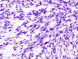
A microscopic image of Leiomyosarcoma cells.
Leiomyosarcoma belongs to a rare class of cancers called soft tissue sarcomas.
Sarcomas arise in connective tissue, which is found all over the body. As a wide variety of bodily structures are made of soft tissue, including muscle, fat, nerves, and blood vessels, there are many potential areas that can harbor leiomyosarcoma. However, its most common occurrence site is the uterus.
Soft tissue sarcomas are exceedingly rare, only comprising 0.7% of all cancer cases, and leiomyosarcoma is rarer still, only accounting for 5-10% of these. However, it can still be considered a serious threat due to several factors, including the difficulty of diagnosis and the aggressive and treatment-resistant nature of the disease.
Specific causes for the disease have not yet been established and thus advice for prevention is limited. An in-depth discussion of Leiomyosarcoma, compiled by the Liddy Shriver Sarcoma Initiative, can be found here.
Any cancer diagnosis is best performed with a variety of information sources, including the patient’s personal health history and family history of disease, risk factor assessment, physical examination, radiological imaging such as MRI, CAT, or PET scanning, and pathological testing on biopsied samples.
However, there are some features of leiomyosarcoma that can make diagnosis particularly challenging.
Some risk factors for uterine leiomyosarcoma include obesity and greater body fat distribution in the upper body, early onset of menstruation or late menopause, and a history of breast cancer treatment via the drug tamoxifen.
To aid in the diagnosis, commonly-used imaging techniques can provide comprehensive cross-sectional images of internal organs or possible tumors. Both Computed Tomography (CT) and Magnetic Resonance Imaging (MRI) scans are well-suited for such imaging of soft tissue, though each have specific benefits and drawbacks.
Another imaging method that is less popular but that can sometimes offer greater sensitivity in detecting tumors is the Positron Emission Tomography (PET), which produces a different type of image (based on physiologic activity) than the other methods. More information on imaging techniques used in uterine cancer diagnosis can be found from the American Society of Clinical Oncology.
Unfortunately, in the case of leiomyosarcoma, imaging techniques cannot generally offer conclusive evidence. This is because uterine leiomyosarcoma tumors, when viewed with such techniques, have a very similar appearance to uterine fibroids (medically termed leiomyomas), which are benign growths in the uterine wall.
Furthermore, uterine fibroids are extremely common—the percentage of women that develop fibroids by age 50 could be as high as 80%, according to the U.S. Department of Health and Human Services.
This is why, according to Martee L. Hensley, MD, MSc, who is an expert in gynecological oncology, there is currently no definite way to diagnose uterine leiomyosarcoma by non-surgical means.
The only reliable method of detection requires testing of a surgically-obtained biopsy by a pathologist experienced with gynecological cancers. Cell samples from the biopsy are stained with pigments for visual contrast for examination via microscopic techniques, and specific features are interpreted in a pathology report.
For example, a sample of leiomyosarcoma tumor is more likely to have a high number of unusually large cells compared to a sample of benign tissue. However, as explained here, taking biopsies before uterine surgery to screen for leiomyosarcoma is not a viable approach, and if a morcellator is used in the surgery, the patient may have already been exposed to the considerable risks.
Treatment approach in a cancer case depends on a wide variety of factors, including the stage (degree of spread) of the cancer and the grade (degree to which the tumor resembles normal tissue) of the tumors, as well as other circumstances surrounding the patient.
The National Cancer Institute has a comprehensive resource page regarding uterine sarcoma treatments.
Developing a treatment plan for a Leiomyosarcoma patient is a complex endeavor, often requiring the expertise of a multi-disciplinary team of specialists. Some common treatment methods are as follows:
Sadly, as leiomyosarcoma is difficult to detect and is resistant to several of these treatment methods, as well as being notoriously aggressive, survival rates for the cancer are low, even with early detection.
The lethal nature of this type of cancer is a truly horrifying aspect of the power morcellator controversy, which is why many patients and families have sought out personal injury attorneys to file suit and gain some sort of compensation.
A number of patients, after undergoing laparoscopic uterine surgeries, which they perceived to be a relatively safe alternative to open surgery, found themselves faced with advanced leiomyosarcoma and possibly robbed of years of life.
If you or your loved one may have been negatively affected by power morcellators during uterine surgery, do not hesitate to contact the New York law firm, Banville Law for a free case evaluation.
To see Settlements For Power Morcellator Cases, see our article.